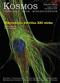
New dimension in microscopy - laser scanning confocal microscope
Abstract
Laser scanning confocal microscope is an invaluable tool for a wide range of research in the field of biological and medical sciences. Confocal microscopy allows to create a thin, optical cross-sections of live or fixed specimens. This characteristic allows for a more precise observation of the structure on the examined sample as well as creation of three-dimensional (3D) image reconstruction. This is possible thanks to the confocal aperture (the pinhole), which passes to the detector only light which comes from the focal plane, so from the layer which has the best optical performance. Modern confocal systems use lasers, which are point-light sources, to excite fluorescent dyes present in the specimen, and point detectors for analysis of the emitted fluorescence. Continuous improvement of confocal microscopes allows to achieve: better resolution of generated images (resolution below 35 nm in the XY axis), greater sensitivity of light detectors (even detection of single photons) and increasingly rapid visualization of samples (scanning speed up to tens images per second). With these enhancements, laser scanning confocal microscopy has gained tremendous popularity in the research community.Downloads
Download data is not yet available.
Downloads
Published
2017-12-09
Issue
Section
Articles


